Last edit:
05-08-30 Graham
Wideman |
Tools
|
GW Raw MRI View
Article created: 2003-06-22 |
(Formerly GWORC: GW Orientation Reality Check)
Overview
GWRawMRIView is a viewer for MRI files that is particularly suited to showing
how the raw data is organized without trying to be too helpful. It is guaranteed
not to understand any particular format or headers.
Update 2004-10-20
I've been using this tool regularly and have added a few features along the
way, and have only now got around to posting a new version. See below for
details.
Why?
This tool is especially useful if you are trying to determine how your raw
MRI data is ordered, independent of what the header might say, and without
confusion that comes from a viewer attempting to show a desirable orientation.
GWRawMRIView shows several images that are clearly marked as to how the images
relate to data order in the data file.
These kinds of issues arise when converting between different file formats,
and when using Analyze-format data or the
newer NIFTI format, the BRIK data files of AFNI, raw MRI scanner files, and also
2-D slice images of various kinds.
System Requirements
- Runs on Microsoft Windows
- Works better with >= 1280 x 1024 display, but that's not required.
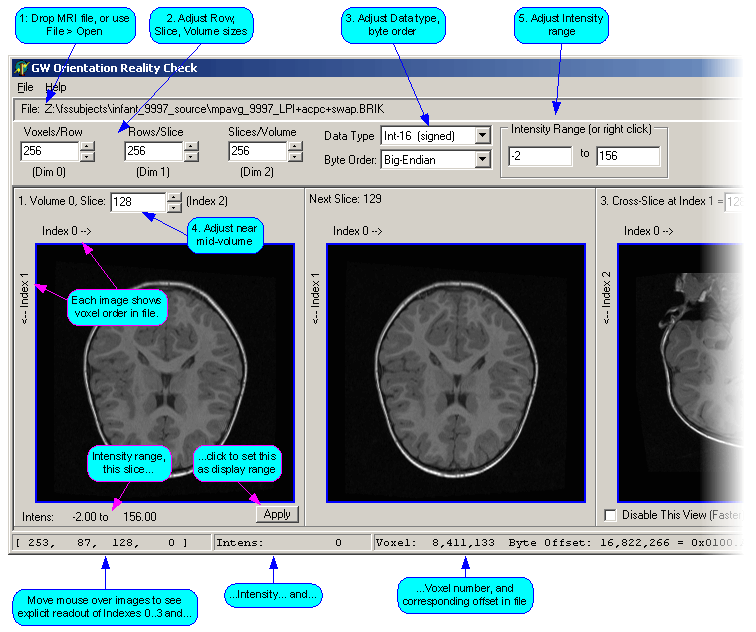
Screenshot
(This is of version 0.1.0.0 -- a few more features have been added, details
below.)

Features Not Appearing in Above Screenshot
- Over/Under checkbox. This cause the display to color voxels that
are above (red) or below (green) the selected intensity range.
- Skip Header Setting: A text box where you enter a number of
initial bytes to skip, typically to skip over a header.
- File > Save Image 1: This feature allows you to save the image in
the first panel as a BMP file.
Installation
- Use link below to download zip file.
- Unzip and place in some convenient directory. No further installation
required.
- Optionally drag to Start menu to create menu item, or Ctrl-Shift-Drag to
desktop to create shortcut.
Use
- To run, double click in Windows Explorer.
- Drag an MRI file or 2D raw image file from Windows Explorer onto File-path
area.
- While observing resulting images, adjust the following in any order:
- Voxels/Row
- Rows/Slice
- Slices/Volume
- Data type and byte-order
- Volume 0 slice number
- Intensity range
- Readings: In addition to the images themselves, you can also view various
readings in the status bar area
Hints
- You will probably want to adjust the Volume 0 slice number (leftmost
image) to somewhere near mid-volume, as the outer slices are usually
all-black. Same with the Index 1 setting for the Cross-slice image.
- Indexes 0, 1, 2, 3: Index 0 is the fastest varying index, then Index 1 and
so on. These correspond to position-in-row, row-in-slice, slice-in-volume, and
volume-in-file. (You could also think of them as X, Y, Z, Vol, so long as you
don't attribute any special spatial meaning to these letters!)
- There's a label showing direction of increasing index for each one, so for
example Index 0 increases from left to right in Images 1 and 2.
- As you mentally correlate the images with how the bytes are ordered in the
file, you can drag the mouse over an image to see how Voxel number and Byte
Offset vary.
Download
| Item |
Version |
Date |
Link |
Description |
| GW Raw MRI View |
0.3.0.0 |
2004-10-20 |
GWRawMRIView_0300.zip |
Adds Header Skip and Save Image features |
| GWORC |
0.1.1.0 |
2003-11-11 |
GWORC0110.zip |
Adds ability to flag in color voxels whose intensity is
outside set intensity range |
| GWORC |
0.1.0.0 |
2003-06-22 |
GWORC0100.zip |
|
| |
|
|
|
|
Go to:

