Last edit: 05-08-29 Graham Wideman
|
Last edit: 05-08-29 Graham Wideman |
FreeSurfer |
| FreeSurfer Surfaces Intersected with MRI Data Article created: 2003-03-10 |
This page provides some samples showing FreeSurfer-produced "final" pial and white surfaces intersected with the MRI data that produced them.
The surfaces are, of course, 3D and in viewing them this way some effort is required to visualize whether the surface at any particular point is relatively perpendicular to the image, relatively parallel, or some angle in between.
Of particular note: FreeSurfer has produced surfaces, and thus outlines, which are not limited to incremental voxel coordinates -- the result is thus finer than the voxel resolution.
| Pial (red) and white
(yellow) surfaces intersected with orig (ie:averaged) coronal MRI slices 126
through 130 (ie: mid volume). FreeSurfer currently only works with volumes that are 256 mm cubes, with 1 mm voxels.
|
126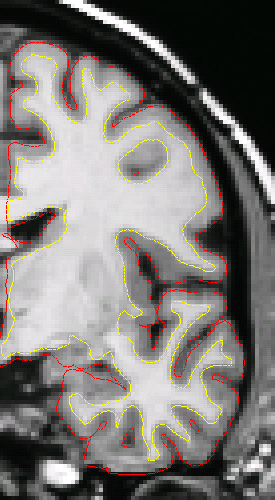 |
127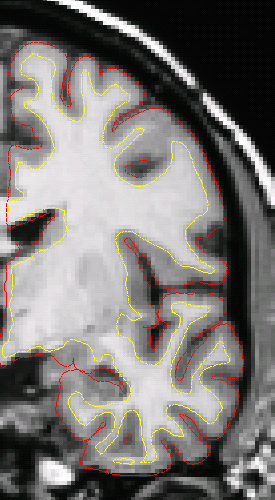 |
128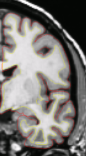 |
129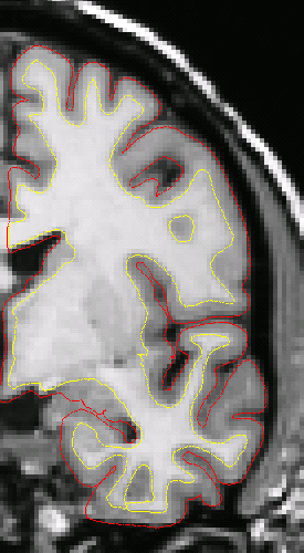 |
130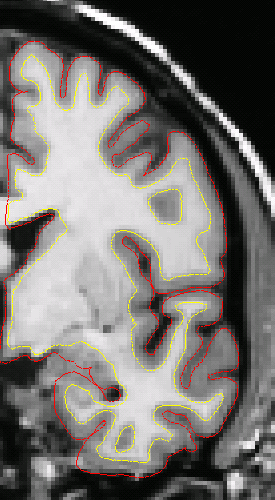 |
| The images here repeat the same slices as above, but this time plotted on the "brain" volume -- after intensity normalization and skull stripping. This gives a sense as to how the skull strip does or does not encroach upon the pial surface. | 126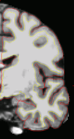 |
127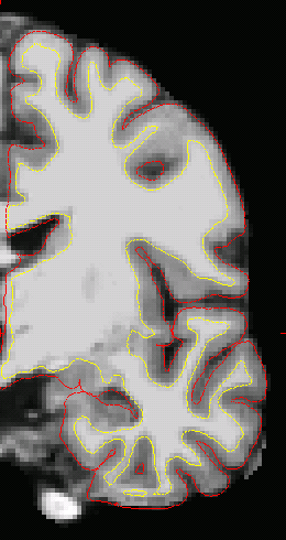 |
128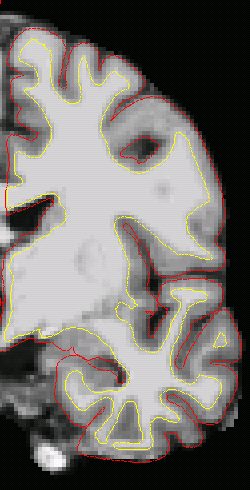 |
129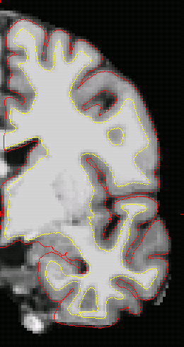 |
130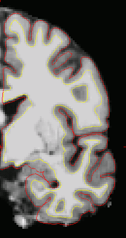 |
Go to:
![]() or back to Understanding FreeSurfer
or back to Understanding FreeSurfer